Low Temperature, Ultra-High Vacuum STM
Device Specifications
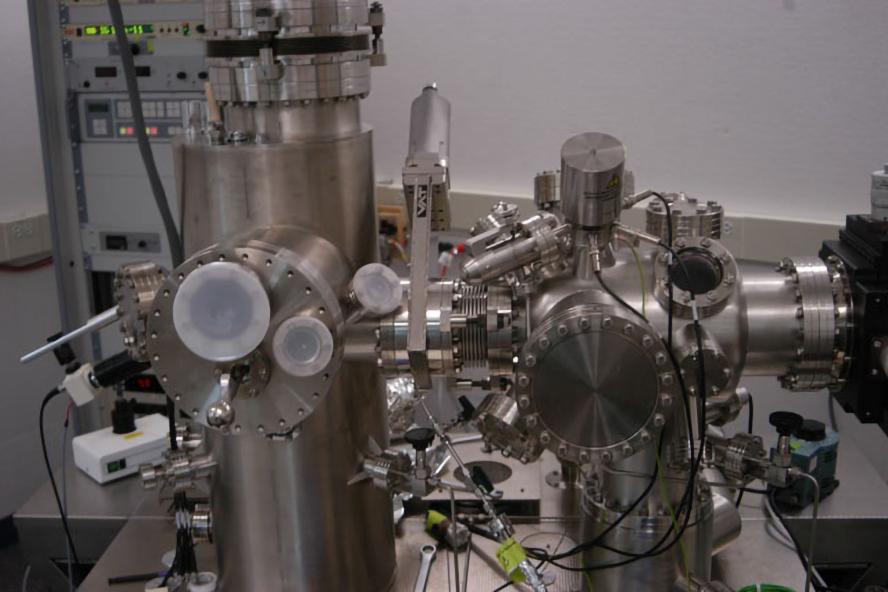
Omicron ultra-high vacuum low temperature STM Setup
STM Chamber
- Lowest Temperature: 5.1 K
- Cooldown from Ambient to 5.1 K: 6 hours
- Time between Helium fills: ~20 hours
- Z resolution: 0.005 nm
- Gap Voltage: +/- .5 mV to 10 V
- Tunneling Current: 2 pA to 50 nA
- Pressure: down to 1x10-11 mbar
Prep Chamber
- Argon Cation Sputter Gun
- Resistance Sample Heater
Capabilities
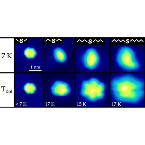
Figure 1: STM images showing thermal activation of thioether rotors; dimethyl, diethyl, dibutyl and dihexyl sulfide. Temperatures in the bottom row indicate the onset of rotation. Tip bias = -0.3V I = 9 pA
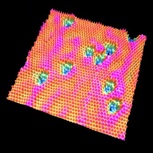
Figure 2: Atomically resolved 3-D STM image of Pd/Cu{111} at 80 K. The Pd atoms were deposited at a sample temperature of 1020 K, providing enough thermal energy for the Pd atoms to bury beneath the Cu surface. The blue depressions represent Pd impurities in a Cu{111} lattice. Electron standing waves appear as pink waves across the surface and around the subsurface Pd atoms. Image conditions: 0.05 V, 0.2 nA, 10 nm x 10.8 nm.
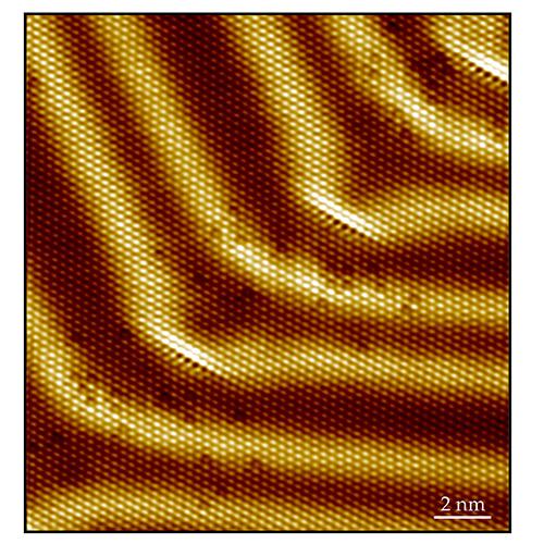
Figure 3: Atomically resolved STM image of Pd/Au{111} at 7 K. The Pd atoms were deposited at a sample temperature of 377 K. Pd atoms place exchange with Au atoms at the edge dislocation of the herringbone reconstruction, alloying into the surface. The edge dislocation appears as a bright line of atoms at the herringbone elbows, while Pd atoms appear as depressions. Image conditions: 0.01 V, 1.3 nA.